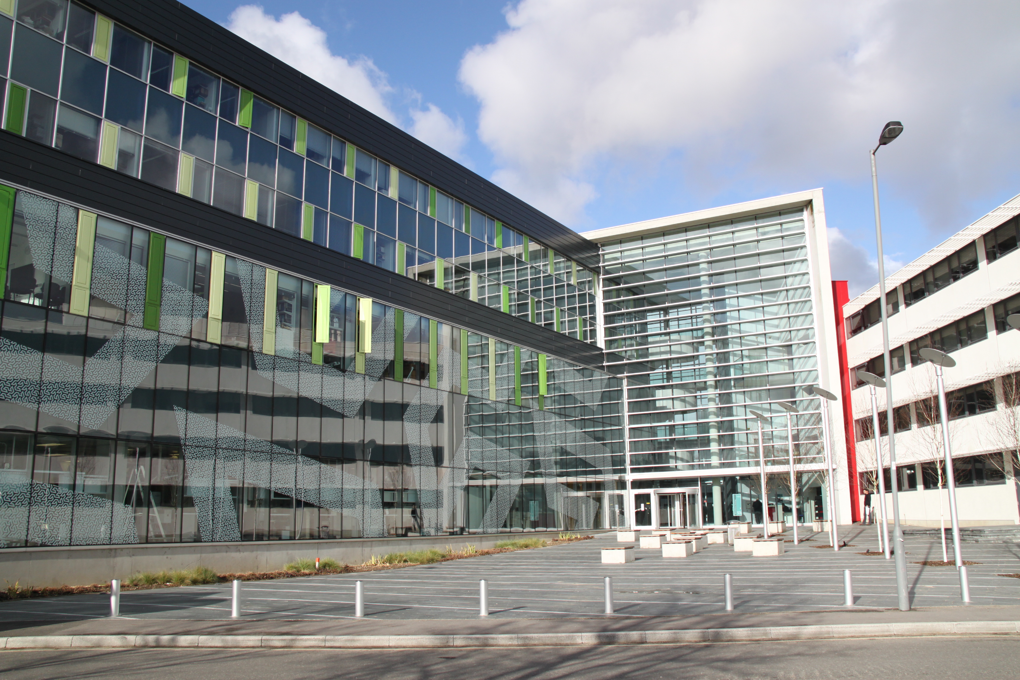
New Mountbatten Photo
The clean room provides a range of characterisation equipment for device and material characterisation. Plan view imaging of a sample surface can be performed using field emission scanning electron microscopy (FESEM), helium ion microscopy and scanning probe microscopy. Cross-section imaging can be achieved by first making a cross-sectional cut using the focussed ion beam (FIB) system and then imaging in-situ using field emission scanning electron microscopy.
DE GROOT, CORNELIS -  +442380592732
You can download the raw data used to create this page:
The following open datasets were used to build this page:
© 2025 University of Southampton

